2026: Volume 7, Issue 1
Current Issue
 Abstract
  PDF
PDFSensing of Heavy Metals by Jasmine Camelia Sinensis Stabilized Copper Nanoparticles
Sheu Sikirat Kehinde
University of York, United Kingdom
*Corresponding author: Sheu Sikirat Kehinde, University of York, United Kingdom, Phone: 07795493080, E-mail: [email protected]
Received: May 29, 2026 Published: June 15, 2026
Citation: Kehinde SS. (2026). Sensing of Heavy Metals by Jasmine Camelia Sinensis Stabilized Copper Nanoparticles. Nanoparticle. 7(1):22.
Copyright: Kehinde SS. © (2026).
ABSTRACT
Copper nanoparticles were synthesized using green tea (Camellia sinensis ) extracts and the application were channelled towards the sensing of heavy metals. The synthesized green tea copper nanoparticles were characterized using Fourier transform infrared spectroscopy, UV/visible spectroscopy, thermal analysis and powder x-ray diffraction techniques, the results from the UV-visible spectroscopy reveals the existence of localised surface plasmon resonance appeared at 400nm, the synthesized copper nanoparticles exhibit Face centred cubic structure of Copper nanoparticles a (JCPDS-04-0836 )s indicated by the powder X-ray diffraction techniques the detection of silver and gold by green tea copper nanoparticles shows a bathochromic shift towards the heavy metals sensed.
Keywords: Copper Nanoparticles, UV-visible spectroscopy, Camelia Sinensis
INTRODUCTION
The greener route of fabricating a metal nanoparticle is a new field at the intersection of nanotechnology sustainable ways that proffers a safe and clean ways of synthesising metal nanoparticles and potentially offers a lasting and promising solutions to the problems of environmental and health concerns acquainted with the classical mode of synthesising metal nanoparticles. Due to the increase in the environmental impacts of traditional ways of synthesizing metal nanoparticles awareness, this calls for a needs for developments of eco-friendly and benign ways for the fabricating of metal nanoparticles. Greener ways of synthesizing metal nanoparticles involve the use of safe and eco-friendly approach to produce the metal nanoparticles. the unique significance of greener ways of synthesizing metal nanoparticles solely lies in the ability to revolutionize different sectors and proffer a sustainable approach to solve global challenges [1,2].
Nanoparticles can be produced using different techniques which includes sol gelling, electrodeposition [3] and sonication [4], however these synthetic techniques are not cost effectives and also lead to production of secondary pollutant to the environments, Greener routes of synthesis ways of fabricating a nanoparticle offers a desired alternative which utilizes plant extracts, bacteria, yeast to reduce and stabilize the metal to its nano states. Greener synthetic routes offer a cost friendly approach, environmentally benign and excludes the use of toxic chemicals and addictive to produce the metal nanoparticles. Greener synthetic routes also offer a large scale production of the metal nanoparticles. The process involved in the greener synthetic routes involves the use of the reductants which are usually the bioactive components of the plant extracts ,which reduces the metal ions in the metal salts which acts as the source of the metal ion generally termed a metal precursor and the plant extracts usually play a dual role, the role of reductant and also a stabilizers, after the reducing of the metal to its free state, the surface are usually prevented from further agglomeration, the bioactive compounds present in the extracts also serves as a stabilant that stabilizes the surface of the metal nanoparticle from aggregation. A typical plant extracts process some a wide range of secondary metabolites such as alkaloids, polyphenols, terpenoids and flavonoids which acts as a reductant by donating electron to the metal salts in solution which facilitates the reduction and stabilizing the surface of the nanoparticles [5]. The stabilization of the metal nanoparticles surfaces can also be done by proteins, organic acids which tends to bind to the surface thereby retarding their aggregation. The choice of the stabilizing agents influences the geometry and surface function of the metal nanoparticles [6], Physiochemical parameters such as PH and temperature affect the growth of the metal nanoparticles thereby affecting the geometry and morphology of the synthesized metal nanoparticles [7]. The process of nucleation and growth in greener synthetic routes of reducing the metal ion to the free states involve the use of biologically sensitive components of the plant extract which form the nucleation active centre and serves as a seed permitting growth of the metal nanoparticles. The process of growth involves the simultaneously reducing the metal ion and depositing of the metal ion onto the existing nuclei leading to the extension of the metal nanoparticle, the growth and nucleation rate depend on the metal salt concentration, time and the synthetic condition [8]. The steps involved in the used of plant extracts as a source of fabricating the metal nanoparticles include the boiling in the plant extracts to obtain the bioactive component in the extracts in the aqua form followed by cooling and filtrating in order to remove any sediments in the solution. The aqua extract solution is then mixed with stochiometric quantity of the metal salts, stirred and the changes in the starting colour to final colour is an indicatives of successful synthesis of the metal nanoparticles. Another confirmatory test such as shinning a quartz light through the synthesized metal nanoparticles is another confirmatory test. Leaves, bark roots and different part of a plant can be sourced and used in the metal nanoparticles synthetic process It is crucial to note that the specific mechanisms involved in greener route of synthesizing a metal nanoparticle depends on the plant extract composition, metal precursor, and reaction conditions. Extensive research is needed to evaluate the detailed mechanisms involved in specific systems.
MATERIALS AND METHODS
All chemicals were of analytical grade and were used as obtained from University of York laboratory suppliers. All chemicals and solvents were used as obtained without further purification. The stoichiometry quantities of salts were mixed in deionized water or their respective solvent to prepare the metallic solutions that were used in the experiments. Ahmad Jasmine Green tea bag was source from a friend which gifts as a present. Copper nitrate trihydrate, gold (III) chloride and silver nitrate.
Methods
5 bags of jasmine green tea were placed 1 L round bottom flask containing 1000 ml deionized water and boiled at 120 oC for 3 hours. The extracts were allowed to cool down and the extract was filtered through filter paper-.,10 ml of the aqueous extract was further diluted to 1000 ml deionized water and 0.05 M copper (II) acetate were added to the solution and heated at 90oC for 6 hours. The solution changes from light yellow to brown and the reaction was stopped.6 The copper nanoparticles thus formed were collected by freeze dried method and stored for analyzation. The synthesized GT-CuNPs were characterized using UV-visible spectroscopy, PXRD, SEM, FTIR and TGA, after successful characterization, the GT-CuNPs were applied towards the interaction of heavy metals such as silver and gold.
Heavy metal detection by synthesized green tea metal nanoparticles
The colorimetric measurement of heavy metals such as silver nitrate and gold (III) chloride are carried out by preparing 2-20 ppm solution of each heavy metal. From it 2 ml of each heavy metal concentrations were added to a 20 ml glass vial and 2 ml of the synthesized GT-CuNPs solution were added and left to stand for 5 minutes after which the solution was analysed using UV-visible spectroscopy.
RESULTS AND DISCUSSION
Physical Appearance of Jasmine Green Tea Stabilized Copper Nanoparticles
The colour of the as synthesized Jasmine green tea copper nanoparticles is a definite brownish which is clearly visible to the naked eye (Figure 1).
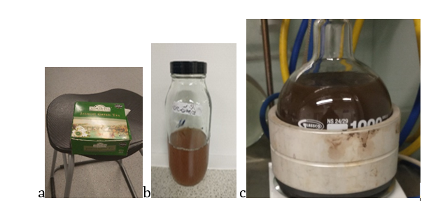
Figure 1: Image of Jasmine green tea bags b) jasmine green tea extract c) jasmine green tea copper nanoparticles
FT-IR of Jasmine Green Tea Stabilized Copper Nanoparticles
The FT-IR of Jasmine green tea extract, there are characteristics spectra of broad -OH- band at 3241 cm-1 which has shifted in the spectra of jasmine green tea copper nanoparticles to 3291cm-1, the carboxyl ion peak at 1612 cm-1has shifted to 1556 cm-1in the spectra of jasmine green tea copper nanoparticles and also the -C-O- shifted from 1025 cm-1 to 1052 cm-1. Depicted in Figure 2 comparing the ascorbic stabilized copper nanoparticles with the jasmine green tea stabilized green tea copper nanoparticles there appears to be presence of more organic component in the ascorbic stabilized copper nanoparticles and trace organic components in the Jasmine green tea copper nanoparticles.
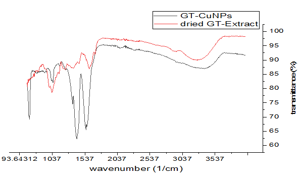
Figure 2: FT-IR spectroscopy of Jasmine green tea extract (dried) and Jasmine green tea copper nanoparticles UV-visible spectroscopy of jasmine green tea stabilized copper nanoparticles
The UV/visible spectroscopy of jasmine green tea stabilized copper nanoparticles were recorded from 200-800nm at a 10 minutes’ interval for 100minutes, the results of the observation are depicted in Figure 4. The LSPR are observed at 412 nm and tends to be stable with time. From the comparison of the UV-vis of spectra of copper acetate, green tea extracts and the GT-CuNPs there exist possible formation of new products (Figure 4). Complete reduction and stabilization of the GT-CuNPs can be observed as there exist a new peak i.e. the LSPR of the Cu-NPs which is absent in the UV-vis spectral of the green tea extracts and the metal salts used in the course of synthesis.
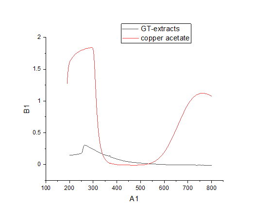
Figure 3: UV/visible spectroscopy of green tea extracts and copper
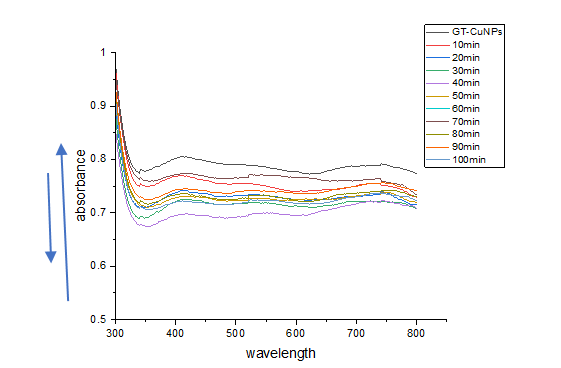
Figure 4: UV/visible spectroscopy of jasmine green tea stabilized copper nanoparticles absorbance stability measurement with time (10-100 mins) at 10 mins interval.
Powder X-Ray Diffraction of Jasmine Green Tea Copper Nanoparticles
The diffractogram peaks at 23.530 (1 1 0), 36.10 (1 1 1), 38.10 (2 0 0),61.460 (2 2 0),75.210 (3 1 1), 48.06o are indexed to copper (II) oxide, the as prepared GT-CuNPs yielded peaks at 45.56˚ (1 1 1), 49.15˚ (2 0 0), 74.38˚ (2 2 0), and 83.49˚ (2 2 2) are indexed to copper, there were some characteristics trace amounts of copper (II) oxide in the diffractogram. (JCPDS No. 04-0784) [26]. XRD spectrum of the synthesized CuNPs product (Figure 5) shows sharp and high characteristic peaks at 2θof (220) planes in standard JCPDS-04-0836 for metallic copper crystals [26]. These peaks confirmed the formation of mainly copper metal in the reaction between Copper acetate and green tea extracts. Some small peaks of Cu2O, CuO, and Cu (OH)2 impurities in the XRD spectrum were possibly due to surface oxidation of metallic copper by oxygen in the air or incomplete reduction of copper (II) by green tea extracts.
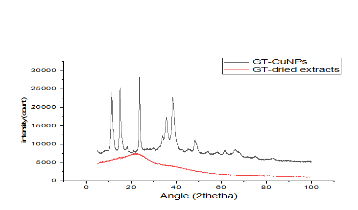
Figure 5: PXRD of Jasmine green tea copper nanoparticles
Thermogravimetry Analysis of Jasmine Green Tea Copper Nanoparticles
The thermogram of GT-CuNPs suggests 32.5 percent’s organic compound presents at the surface of the synthesized GT-CuNPs, the first decomposition is attributed to the loss of water molecules in the sample and the huge decomposition occur between 120-280 0C suggesting the decomposition of the organic components of the green tea. The reduction and stabilization of the CuNPs by the GT extracts contains a high amount organic compound (Figure 6).
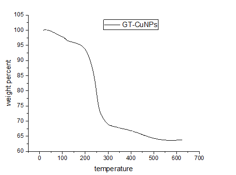
Figure 6: Thermal analysis of GT-CuNPs
Scanning Electron Microscope of Jasmine Green Tea Copper Nanoparticles
The SEM images of GT-CuNPs shows a degree of aggregation in the morphology.? (Figure 7)
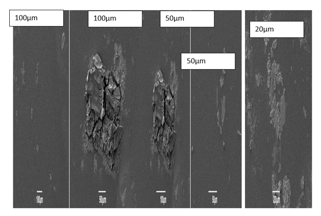
Figure 7: SEM images of GT-CuNPs
Particle size analysis are not carried out due to the unavailability of the instruments in the laboratory and limited time to send it for analysis.
Sensing of Gold by GT CuNPs
The interaction of gold with GT-CuNPs leads to the appearance of precipitates and also the retention of the pinkish colour of the synthesized GT-CuNPs. The LSPR of GT-CuNPs at 415 nm and there is another appearance of second peak at 535 nm of Au (III). The interaction of silver by GT-CuNPs shows the appearance of pinkish brown at different concentration of the silver ions, the LSPR has been observed to shift from 411nm to 415nm, the appearance of the LSPR remains the same even after the complexing of the silver ions with the GT-CuNPs (Figure 8,9).
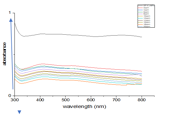
Figure 8: UV-vis spectroscopy of GT-CuNPs and gold (2-20 ppm)
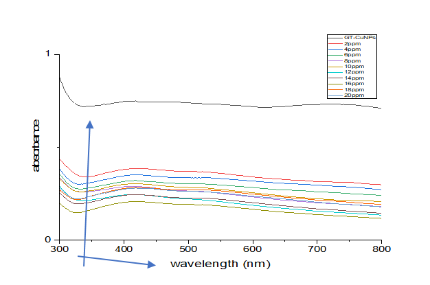
Figure 9: UV-vis spectroscope of GT-CuNPs and silver (2-20 ppm)
CONCLUSION
The report used a benign approach in the fabrication of the copper nanoparticles this approach is environmentally friendly and it avoid the use of toxic or harsh chemicals in the course of fabrication of the nanomaterial. This report shows the potential of jasmine green tea extract to reduce copper atoms to its zerovalent state, results from various characterization techniques such as UV/visible spectroscopy reveals the existence of localized surface plasmon resonance and the localized surface plasmon resonance tends to be stable with time examined ,the synthesized jasmine green tea stabilized copper nanoparticles exhibits a face cantered cubic structure, the thermogravimetric analysis reveals the existence of 32.5% organic compound with 67.5% copper and remnant. The application also reveals the potential ability of the synthesized jasmine green tea stabilized copper nanoparticles to be used a detection tool for detecting heavy metals from waste water. As they are extensively used in detection tools like biosensors and colorimetric assays for environmental monitoring, food safety, and medical diagnostic.
REFERENCES
- Vijayaram S, Razafindralambo H, Sun YZ, Vasantharaj S, Ghafarifarsani H, Hoseinifar SH, et al. (2022). Applications of Green Synthesized Metal Nanoparticles — a Review. Environmental Research. 202(1):360-386.
- Kumari S, Raturi S, Kulshrestha S, Chauhan K, S.Dhingra S, András K, et al. (2023). Journal of Materials Research and Technology. 27:1739-1763.
- Mkawi EM, Ibrahim K, Ali MKM, Farrukh MA, Mohamed AS. (2013). Dependence of Copper Concentration on the Properties of Cu2ZnSnS4 Thin Films Prepared by Electrochemical Method. Trans Tech Publ. 343:85.
- Gandlevskiy N, Viana AR, Druzian GT, Oliveira DK, Schuch AP, Barge A. (2025). Ultrasound-assisted green synthesis of silver nanoparticles using Ruta graveolens L. Extract and antitumor evaluation. Ultrasonic sonochemistry. 117:107340
- Hanna DH, Nady DS, Wasef MW, Mohamed MHFHS, Isaac DM, Kirolos MM, et al. (2025). Plant-derived nanoparticles: Green synthesis, factors, and bioactivities. Next Materials. 9:101275.
- Piergies N, Oćwieja MC, Paluszkiewicz C, Kwiatek WM. (2025). Nanoparticle stabilizer as a determining factor of the drug/gold surface interaction: SERS and AFM-SEIRA studies. Applied Surface Science. 537:147897.
- Gholipour B, Shojaei S, Rostamnia S, Jamal MRN, Kim D, Kavetskyy T, et al. (2021). Metal-free nanostructured catalysts: sustainable driving forces for organic transformations. Green Chemistry. 23:6223-6272.
- Belloni J, Marignier J.-L, Mostafav M. (2020). Mechanisms of metal nanoparticles nucleation and growth studied by radiolysis. Radiation Physics and Chemistry. 169:107952.
